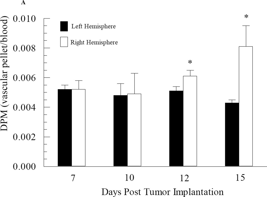
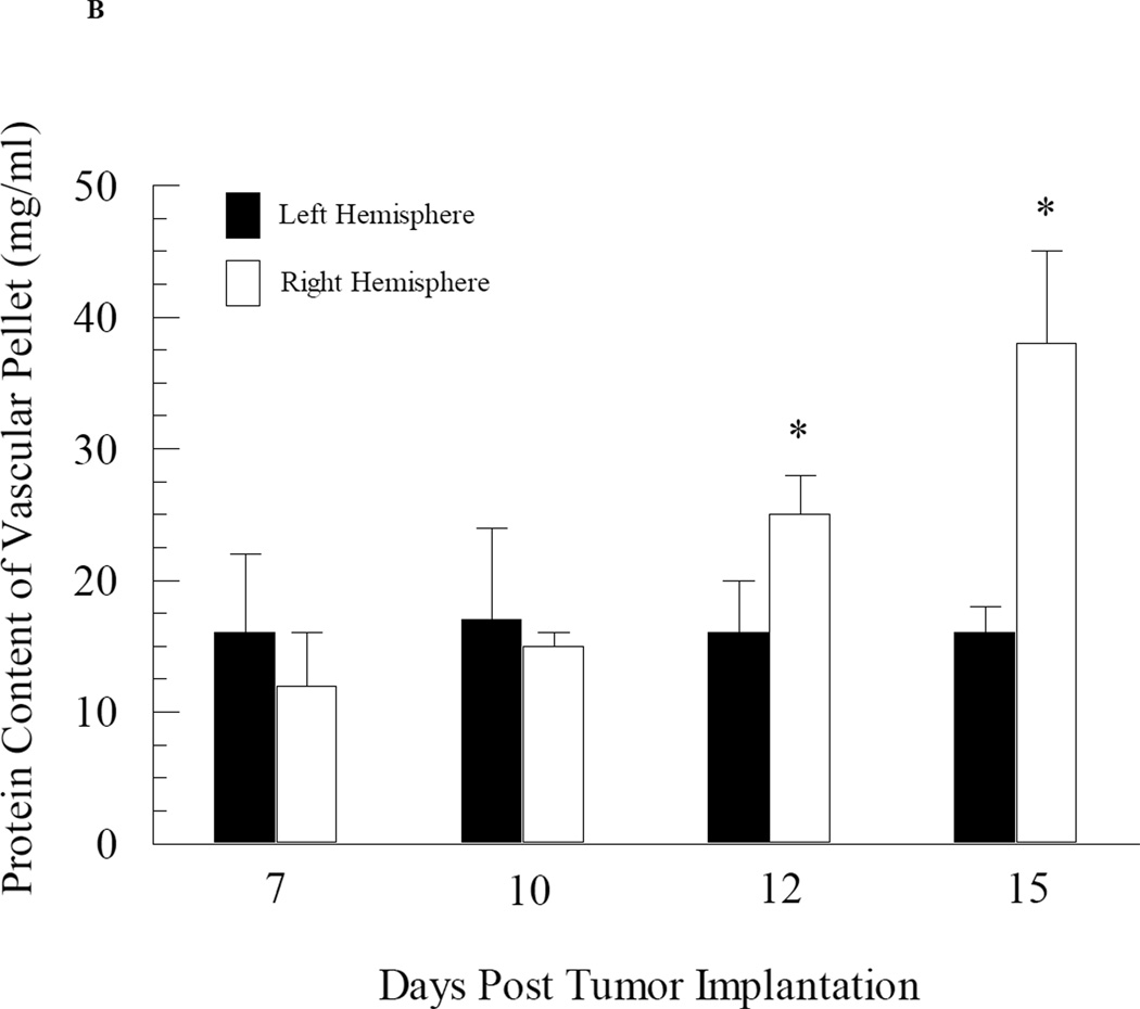
Fig. 3.
Changes in cerebral vascular volume in mice as a function of brain tumor development. Mice at different stages of brain tumor development were given i.v. injections of radiolabelled mannitol. The brains were removed and separated into right (tumor-bearing) and left (non-tumor bearing) hemispheres and capillary depletion was performed. Vascular volume was assessed based on radioactivity (Panel A) and protein content (Panel B) of the capillary pellet. Values represent the mean ± SEM of 6 mice. * p < 0.05 compared to left hemisphere at same day as assessed using Student paired t-test.


