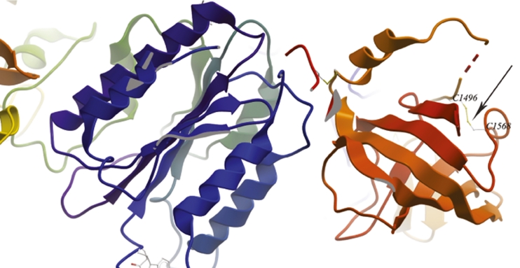
Fig. 3.
Details of the structure of C345C domain of C3 and factor B based on the PDB model 2WIN. The figure shows the C345C domain on the right hand side (in red/orange) and factor B on the left hand side (in blue/green). Note that, in the PDB model, the residues Cys1518 and Cys1590 correspond to Cys1496 and Cys 1568, respectively. The arrow points the disulphide bridge formed by both cysteine residues

