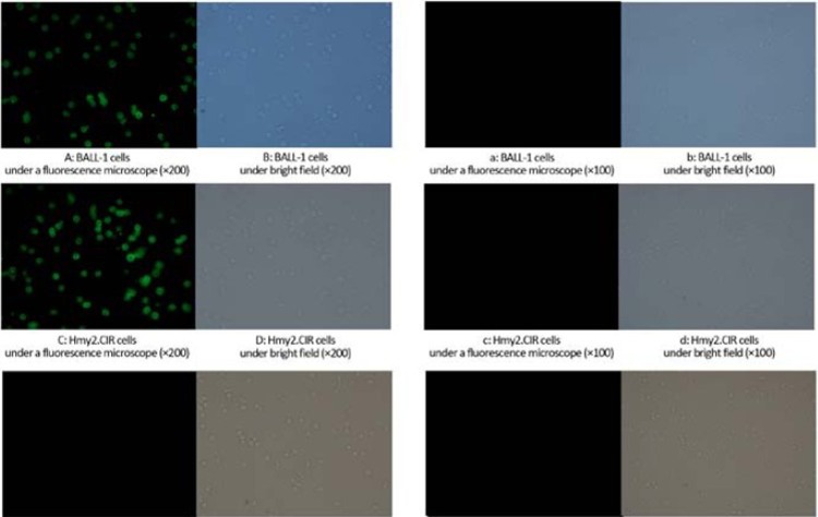
Figure 8.
Cell binding activity of recom-binant protein. A–F: Cell binding activity of fluorescent-labeled recombinant protein; a–f: Cell binding activity of fluorescent-labeled free diphtheria toxin. Under the inverted fluorescence micros-cope, strong green fluorescence could be observed in BALL-1 (Figure 8A) and Hmy2.CIR (Figure 8C) cells with fluorescent-labeled recombinant protein (P<0.05), whereas the negative control group did not show fluorescence in U937 cells (Figure 8E, P<0.05) and fluorescent-labeled free diphtheria toxin BALL-1 (Figure 8a, P<0.05), Hmy2.CIR (Figure 8c, P<0.05) and U937 cells (Figure 8e, P<0.05), indicating that there was a specific binding ability between the recombinant protein and surface receptors on BALL-1 and Hmy2.CIR cells.

