
Figure 1.
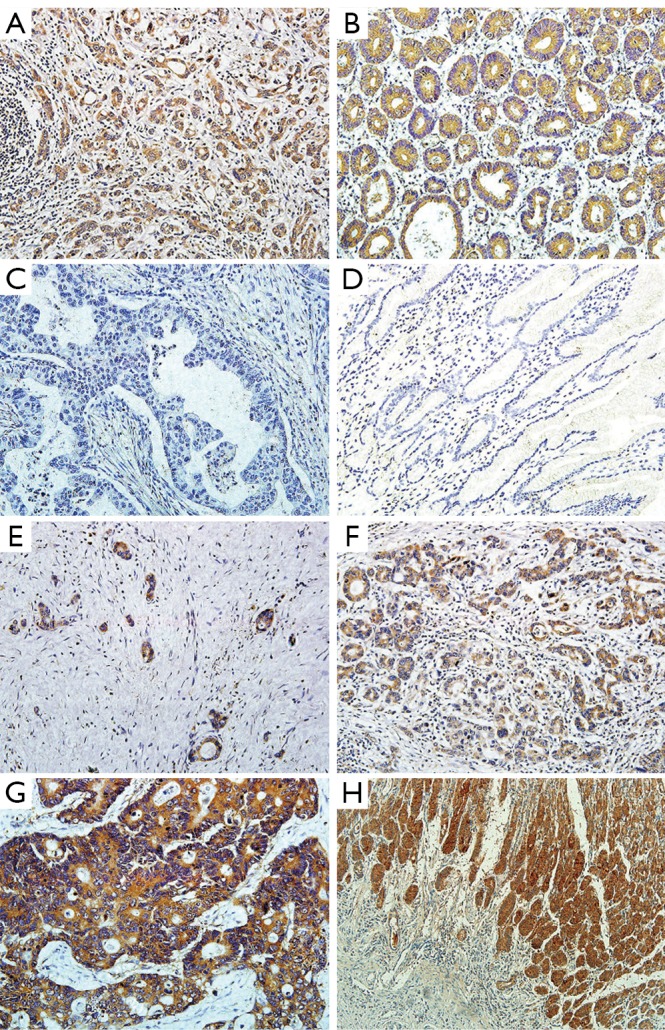
Representative immunohistochemical staining of LGR5 in gastric cancer and adjacent normal tissues. A and B show LGR5 positive staining in gastric cancer and adjacent normal tissues, respectively (magnification ×200). C and D show LGR5 negative staining in gastric cancer and adjacent normal tissues, respectively (magnification ×200). E, F and G show LGR5 staining in gastric cancer tissues with weak, moderate and strong expression (magnification ×200). H shows that LGR5+ cells are present at the base of normal human gastric crypts (magnification ×100)