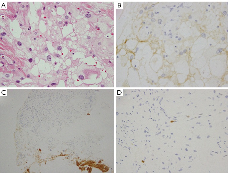
Figure 2.
Histopathological findings of case 10. A. Tumor tissue contained large fat-like proliferation with conventional meningothelial neoplastic cells. Tumor cells have round nuclei and fat vacuole in cytoplasm in case 10 (H&E, original magnification ×40); B. The tumor cells were positive for EMA (original magnification ×40); C. The tumor cells were negative for GFAP (original magnification ×20); D. MIB-1 labeling index was less than 1% (original magnification ×40)

