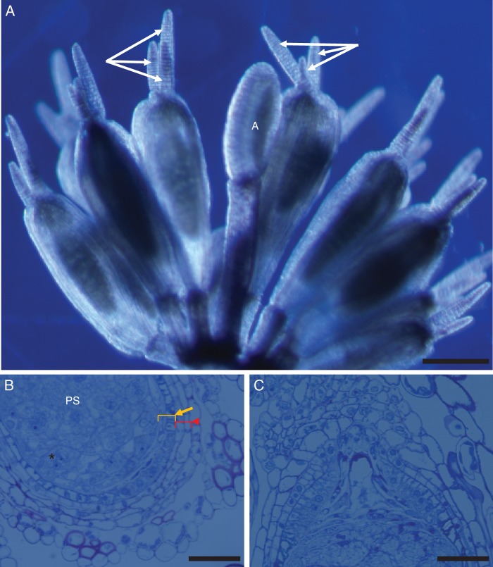
Fig. 1.
(A) Trithuria submersa reproductive unit, with a single stamen in the centre (labelled ‘A’) surrounded by several pistils, each with several long, multicellular uniseriate stigmatic hairs (arrows); scale bar = 200 µm. (B) Detail of lower part of ovule and carpel wall showing perisperm (PS) (*) with aggregated nuclei and two integuments, each with two cell layers (arrow, inner integument; arrowhead, outer integument); scale bar = 50 µm. (C) Detail of upper part of ovule and carpel wall showing the nucellar beak; scale bar = 50 µm.

