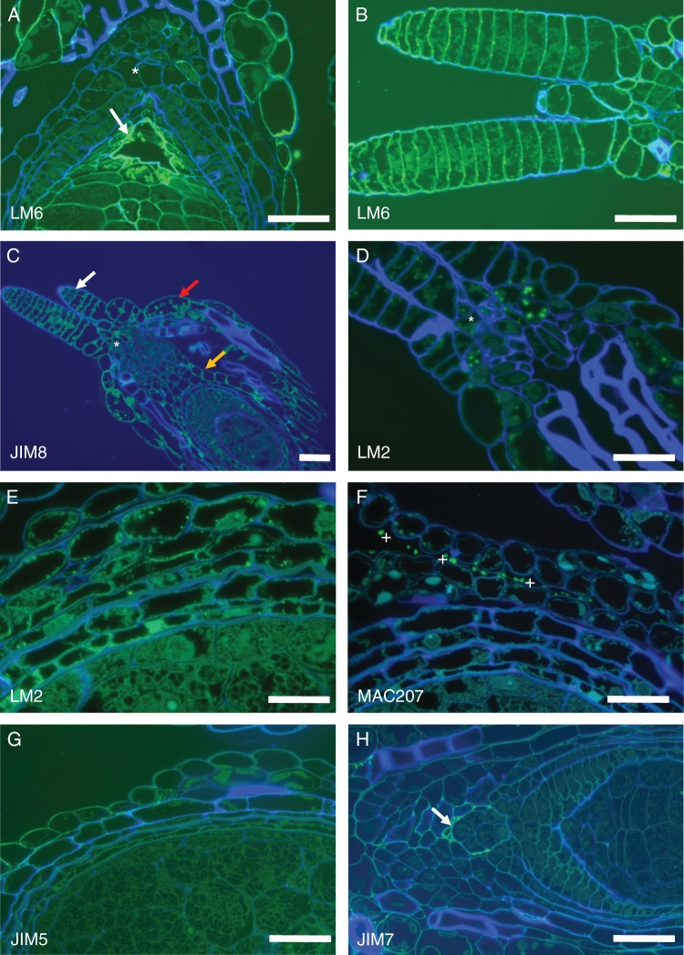
Fig. 3.
(A) Details of LM6 labelling on the embryo sac wall (arrow) and nucellar beak (*). (B) Stigmatic hairs strongly labelled with LM6 which recognizes epitopes in pectins (i.e. the arabinan side-chains) and in AGPs. (C) JIM8 (as well as JIM13) labels the ovary wall (orange arrow), the outer integument (red arrow), the stigmatic hairs (white arrow) and the region at the base of the stigmatic hairs (*). (D) LM2 antibodies intensely label the stigmatic hairs and the ovary entrance (*). (E) LM2-recognized epitopes are strongly present in the integuments and ovary wall. (F) Monoclonal antibody MAC207 consistently labels some secreted material present in the extracellular spaces (*), but not the ovule or the ovary cell walls. (G) JIM5 only labels the outer ovary wall while (H) JIM7 labels the embryo sac wall (arrow) the outer ovary wall and, very faintly, the perisperm. Scale bars: (A, B, D, G, H) = 50 µm; (C) = 100 µm; (E, F) = 20 µm.

