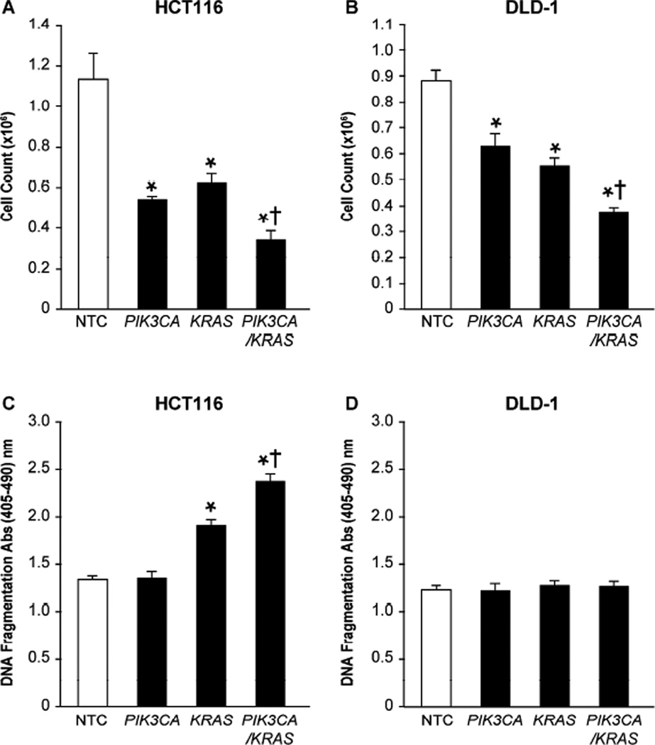
Figure 2. Combined PIK3CA + KRAS siRNA treatments enhance anti-proliferative effects.
(A) HCT116 and (B) DLD-1 cells were plated in 24 well plates at a density of 25,000 cells/well. Cells were transfected 12 h later with 100 nM NTC siRNA, 50 nM PIK3CA siRNA + 50 nM NTC siRNA, 50 nM KRAS siRNA + 50 nM NTC siRNA, or 50 nM PIK3CA siRNA + 50 nM KRAS siRNA. Proliferation was assessed by cell counting 72 h following transfection. (C) HCT116 and (D) DLD-1 cells were plated in 24 well plates at a density of 50,000 cells/well. siRNA transfections were performed using the same treatment groups described above. Media was exchanged for fresh growth media 4 h later. Serum starved conditions were initiated 24 h after transfection and continued for an additional 24 h. Forty-eight h post transfection, apoptosis was measured by DNA fragmentation using Cell Death Detection ELISAplus (* p < 0.05 vs. control; † p < 0.05 vs. PIK3CA siRNA or KRAS siRNA alone).

