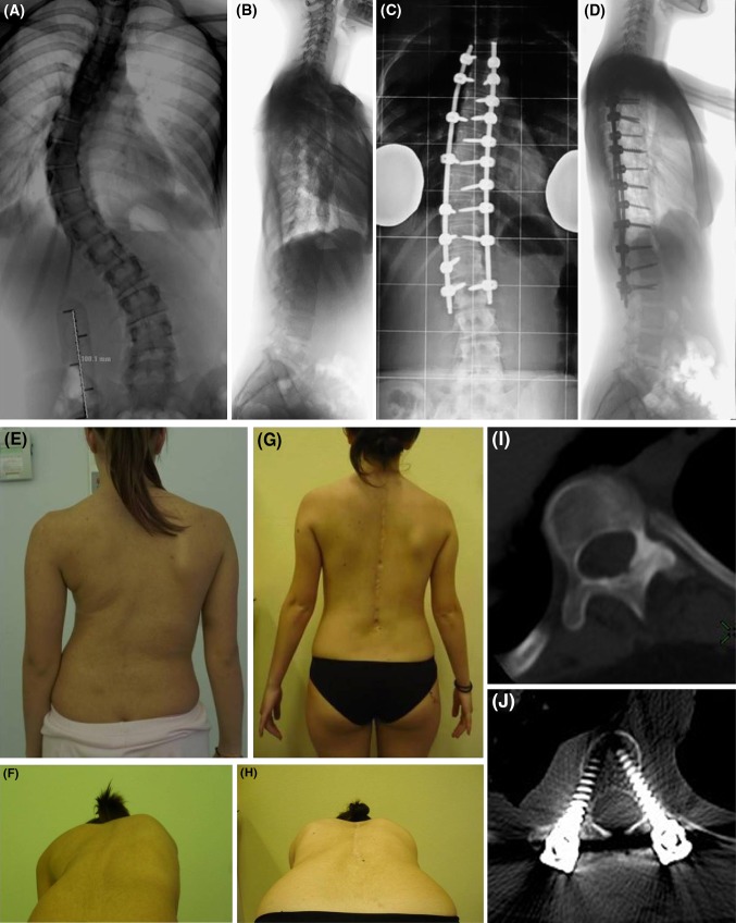
Fig. 2.
DAA, 14-year-old female. AIS: right thoracic (65°) and left lumbar curve (35°), Lenke’s type 1A-curve (a, b). X-rays control 2.8 years after T4–L2 posterior fusion (direct vertebral rotation): the thoracic curve was corrected to 18° (correction of 72.3 %) (c, d). T5–T12 kyphosis remained unchanged at follow-up (8°) (c, d). The clinical aspect before surgery (e, f) and at last control (g, h). Apical RAsag decreased from 29° before surgery (i) to 8° at last follow-up (j) (72 % of correction)

