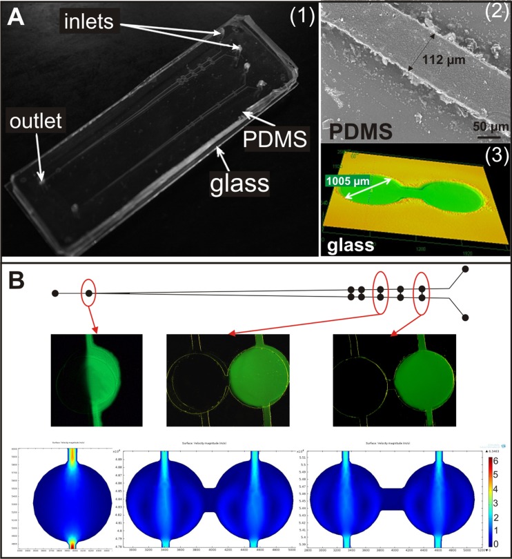
Figure 2.
(a) A photographs of (1) the designed and fabricated microsystem for carcinoma and non-malignant cells migration analysis, (2) the microchannel fabricated in the PDMS plate (Hitachi TM1000), and (3) the microchambers developed in the glass (LEXT OLS3100 Olympus). (b) Tests of solution flow—pairs of microchambers during the introduction of fluorescein solution and distilled water. At the end simulation of flow rate of two introduced substances through microchambers (comsol multiphysics) are presented.

