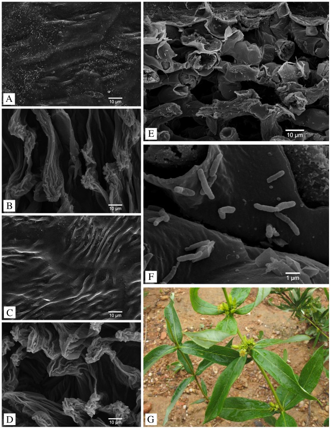
Figure 1. SEM micrographs of the host plant Fadogia homblei.
A. The upper leaf surface before sterilization with a lot of undefined debris. B. The lower leaf surface before sterilization with some debris present. C. After sterilization the upper leaf surface is almost entirely devoid of particles and no epiphytes are visible. D. On the lower leaf surface there are no epiphytes visible after sterilization. E. Cross section through a leaf showing the mesophyll cells with scattered bacterial endophytes. F. Detail of the endophytes in the intercellular space. G. The host plant Fadogia homblei does not have dark bacterial galls on its leaf blades.

