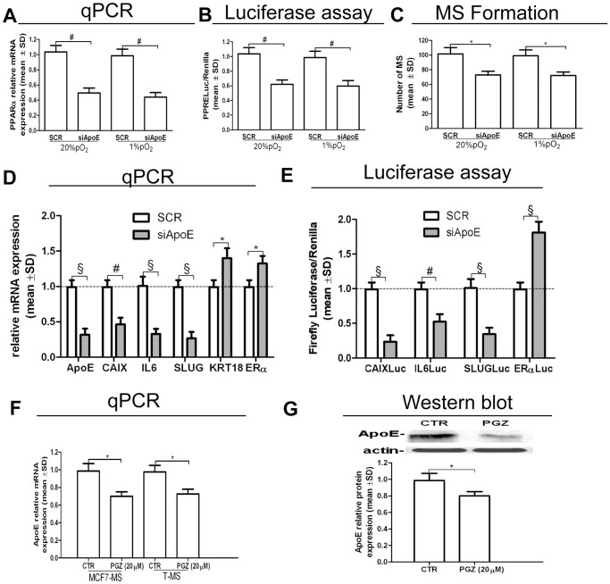
Figure 8. Inhibition of ApoE expression in T-MS.
(A) PPARα mRNA qPCR analysis, (B) PPRELuc activity and (C) MS formation assay in siApoE (48 h)-transfected MCF7 cells, in normoxic and hypoxic condition; (D) ApoE CAIX, IL6, SLUG, KRT18 and ERα mRNA qPCR analysis in siApoE (48 h)-transfected MCF7-MS. (E) CAIXLuc, IL6Luc, SLUGLuc and ERαLuc activity in siApoE (48 h)-transfected MCF7-MS; ApoE mRNA qPCR analysis (F) and ApoE protein expression (G) in PGZ (20 µM, 24 h)-exposed MCF7-MS and T-MS (samples 19–20). Data are expressed as mean ±S.D., n = 3, *p<0.05, # p<0.01, § p<0.005, ANOVA test. n.s.: not significant.

