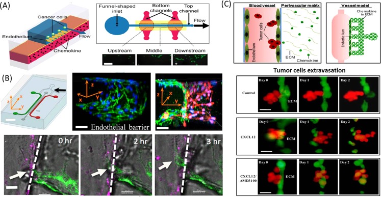
Figure 5.
The process of intravasation and extravasation recapitulated on the biomimetic microfluidic devices. (a) The adhesion of cancer cells on the endothelium layer was region-specifically investigated under physiological flow conditions on the microfluidic vasculature.70 Reprinted with permission from J. W. Song et al., PLoS ONE 4, e5756 (2009). Copyright 2009 Public Library of Science. (b) Microfluidic tumor-vascular interface model for tumor cells intravasation study. The confluent endothelial monolayer can be formed on the 3D ECM. In the process of intravasaton, breast carcinoma cell (white arrow) migrated across the HUVEC mono layer (magenta) in the presence of macrophage.71 Reprinted with permission from I. K. Zervantonakis et al., Proc. Natl Acad. Sci. U.S.A. 109, 13515 (2012). Copyright 2012 National Academy of Sciences. (c) Mimicking the extravasation process of tumor aggregates from the endothelial layer based on the bioengineering blood vessel model.72 Reprinted with permission from Q. Zhang et al., Lab Chip 12, 2837 (2012). Copyright 2012 The Royal Society of Chemistry.

