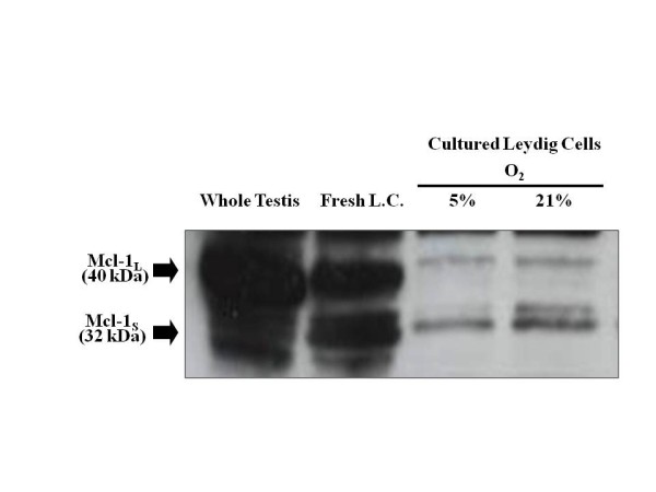
Figure 7.

Immunocytochemical localization of Mcl-1 in the normoxic testis. (A) Negative control section from rat spleen incubated with a goat-anti-rabbit HRP secondary antibody in the absence of the Mcl-1 primary antibody (400×). (B) Positive control section from a spleen section showing Mcl-1 immunoreactivity (400×). (C) Negative control section of normoxic testis incubated with goat-anti-rabbit HRP secondary antibody in the absence of the Mcl-1 primary antibody (400×). (D) Normoxic testis section showing Mcl-1 immunoreactivity in Leydig cells indicated by arrows (400×). The sections were counterstained with hematoxylin. Bar, 100 μm.
