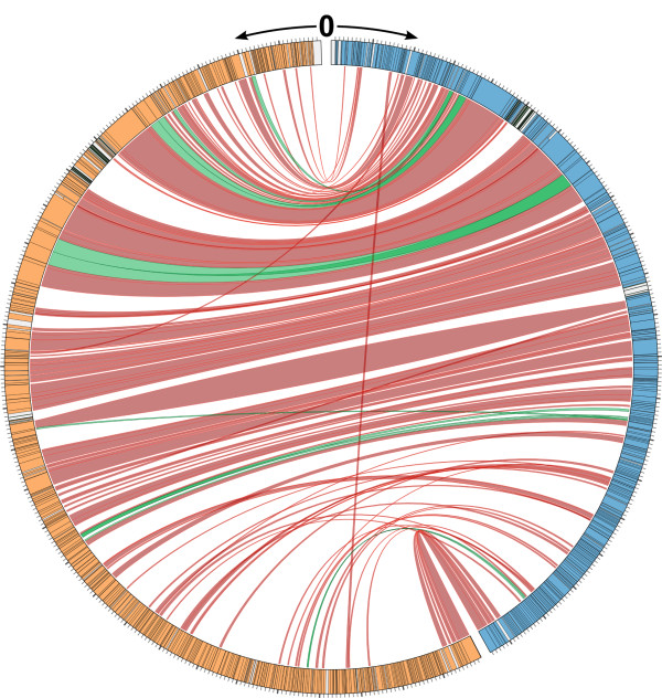
Figure 6.
Map showing the relationship between C. crescentus phages phiCbK and Colossus at the protein level. Both genomes start at position 0 at the top of the figure and move down, with Colossus in orange on the left and phiCbK in blue on the right. Black bands in each track denote the boundaries of protein-coding genes, grey areas represent non-coding regions. Grey tick marks on the outside edge represent 1 kb of sequence, with heavier ticks representing 10 kb. Proteins present in both phages are connected by red ribbons between the two genomes; green ribbons mark proteins with more than one homolog in the other phage, suggesting gene duplications. For clarity, terminal repeat regions were excluded from this figure.

