
Figure 7.
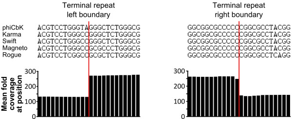
The left and right genomic terminal repeat boundaries of phage phiCbK and four phiCbK-like phages. Terminal boundaries are indicated by the vertical red lines. Above: aligned DNA sequences 12 bp up- and downstream of each terminus are shown; alignments show that the experimentally confirmed boundary sequences of phiCbK are nearly identical to those found in the other four close phiCbK-like relatives. Below: average fold coverage at each base position for all five genomic sequences; note the coverage within the terminal repeats is approximately twofold greater than the surrounding genome, and the breakpoints are identical.