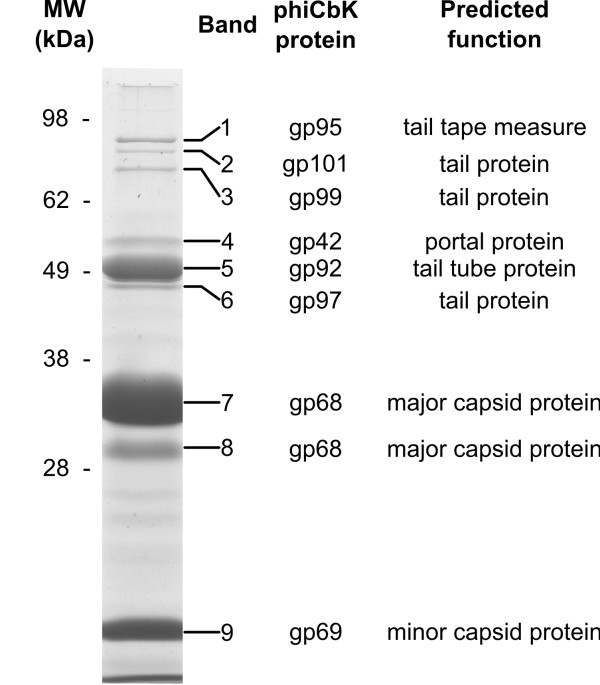
Figure 8.
Coomassie-stained SDS-PAGE gel of purified phiCbK virions. The entire gel lane shown was segmented and subjected to proteomic analysis; band identities and predicted functions are annotated on the right side of the figure. While the entire gel lane was analyzed, only bands that returned conclusive peptide matches are annotated.

