Figure 1.

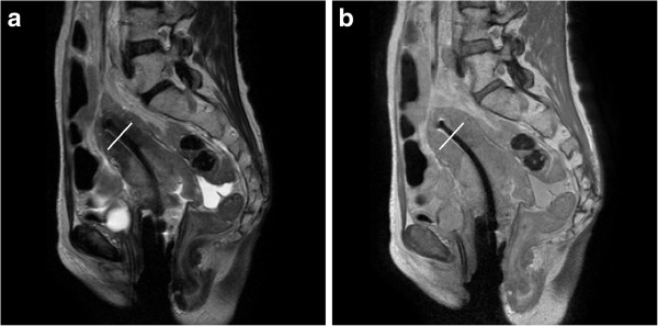
Comparison of (a) T2W and (b) PDW MRI images for localizing tumors and the applicator. White lines across the tandem on both images were added during the post processing to show the location of signal profiles plotted in Figure 2.

Comparison of (a) T2W and (b) PDW MRI images for localizing tumors and the applicator. White lines across the tandem on both images were added during the post processing to show the location of signal profiles plotted in Figure 2.