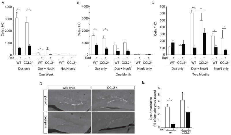
Figure 5. Impaired neuronal maturation following irradiation is partially reversed in CCL2−/− animals.
A–C Confocal evaluation of BrdU-labeled neurons for expression of Dcx and/or NeuN at one week, one month and two months after irradiation (see Fig. 3 for BrdU labeling paradigms). The abundance of immature (Dcx only), transition state (Dcx+NeuN) or mature (NeuN only) neurons was strongly reduced in both WT and CCL2−/− animals at one week (A) and one month (B) following irradiation. Two-way ANOVA, treatment effect, 1 week, F(3,27) = 19.92, p<0.001; 1 month, F(3,33) = 13.87, p<0.001; posthoc Bonferroni, **p < 0.01, *p < 0.05. (C) Cells evaluated at two months following irradiation show that absence of CCL2 significantly increased the number of transition state neurons when comparing CCL2 irradiated versus irradiated WT animals. Two-way ANOVA, F(3,72) = 10.79, p<0.001, posthoc Bonferroni, ***p < 0.001, *p < 0.05. D, E Dcx abundance (pixel area) and staining intensity shows that irradiation results in the depletion of Dcx-positive arbors within the dentate gyrus of WT animals. The absence of CCL2 attenuates this depletion. ANOVA, F(3,32) = 7.690, p<0.001, posthoc Newman-Keuls, *p < 0.05.

