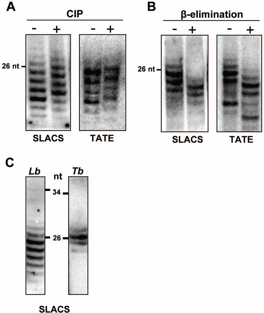
Fig. 1. Structure of L. braziliensis siRNAs.
A. Low molecular weight RNAs were treated with calf intestine phosphatase (CIP) or B. submitted to periodate oxidation/ β-elimination, fractionated on a 15% sequencing gel and analyzed by Northern blotting with SLACS or TATE oligonucleotide probes.
C. L. braziliensis or T. brucei low molecular weight RNAs were fractionated as described above and analyzed by Northern blotting with species-specific SLACS oligonucleotide probes. The samples were run on two separate gels and the lanes aligned using the positions of the molecular weight markers. The positions of relevant 5’ end-labeled pBR322/MspI fragments are indicated.

