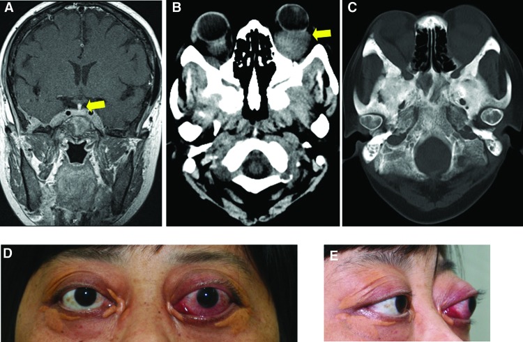
Figure 1.
Exophthalmos and skull images. (A): Pituitary stalk thickening on magnetic resonance imaging (arrow). (B): Orbit computed tomography (CT) showed a granuloma (arrow) in the left retrobulbar space, and bilateral thickening of rectus muscles. (C): Diffuse bone destruction and hyperosteogeny in the skull CT. (D, E): The patient suffered from severe exophthalmos (left, 29 mm; right, 24 mm). Bilateral periorbital xanthomas accompanied the exophthalmos.

