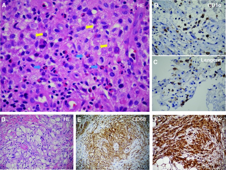
Figure 3.
Immunohistochemistry. (A): Hematoxylin and eosin (HE) staining of the scalp mass showed Langerhans cells (yellow arrows) were distributed in clusters with eosinophils (blue arrows) infiltration. Immunostaining revealed CD1α (B) and Langerin positive Langerhans cells (brown color) (C). HE staining of the retrobulbar mass showed foamy histiocytes nested in fibrosis (D). Immunostaining revealed CD68+ (E) and Lyso+ (F).
Abbreviation: HE, hematoxylin and eosin.

