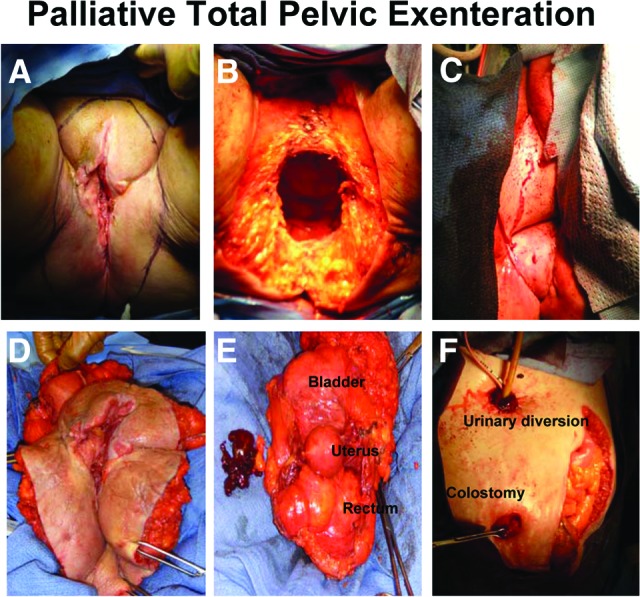
Figure 5.

Palliative total pelvic exenteration. (A): Outline of perineal incision encompassing vulvar lesion. (B): Perineum after removal of pelvic structures. (C): Perineum after reconstruction with ventral rectus abdominus myocutaneous flap. (D): External view of specimen including vulva, vagina, perineal body, and anus. (E): Internal view of specimen including bladder, uterus, and rectum removed en masse. (F): Abdomen after creation of ileal conduit and diverting end colostomy.
