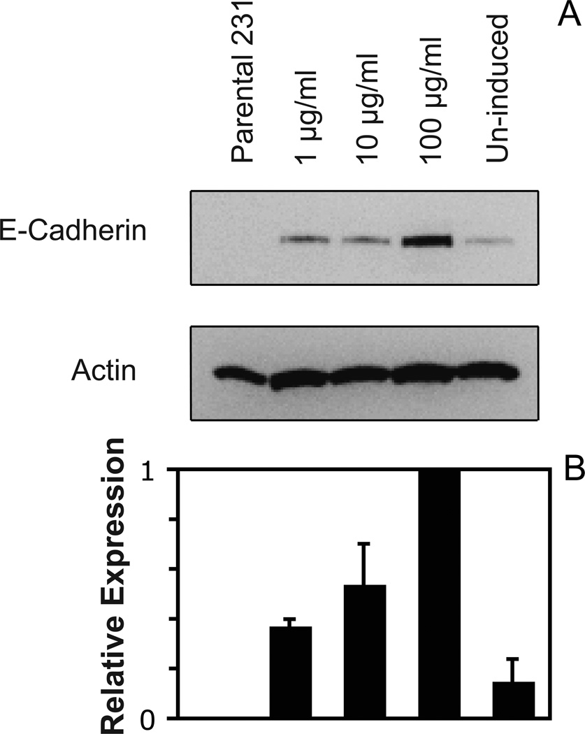
Figure 1.
(A) Western blots of equivalent micrograms of lysates from parental 231 cells and 231-Tet cells treated with different concentrations of doxycyline. The lysates were analyzed using a mouse anti-human E-cadherin mAb (BD Bioscience). The total actin was used as an internal standard. (B) Plot of the E-cadherin expression levels in the different cells in (A), relative to the E-cadherin expressed in cells treated with 100 µg/ml doxycycline. The E-cadherin expression in the different cells was normalized to total actin in the lysate.

