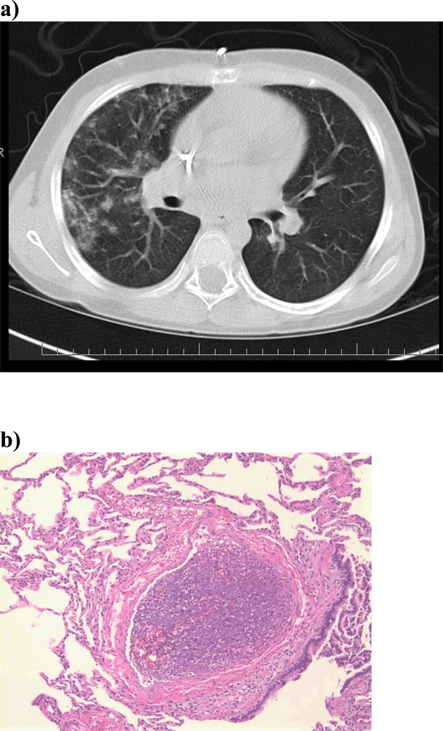
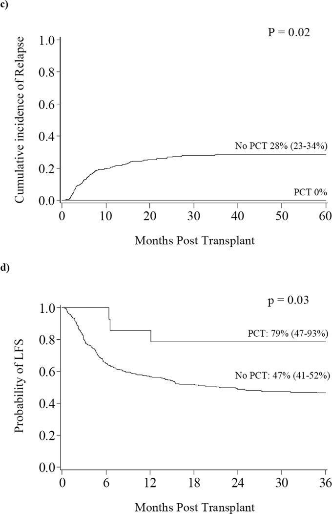
Figure 1. a) Radiographic Appearance of PCT b) Histologic Appearance of PCT c) Relapse d) Leukemia Free Survival.
a) CT scan shows numerous peripheral and pleural based nodular densities in the lung. b) PCT is characterized histologically by necrotic thromboemboli in the distal pulmonary vessels with entrapped leukocytes and amorphous material suggestive of cellular breakdown products. In samples where the cellular debris is still viable, immunohistochemistry shows mainly macrophages (i.e., cells staining for MPO and CD68). c) The cumulative incidence of relapse at three years was significantly lower in those with PCT compared to those without PCT (0% vs. 28%, p=0.02). d) The probability of LFS at three years was significantly higher in those with PCT compared to those without PCT (79% vs. 46%, p=0.03).


