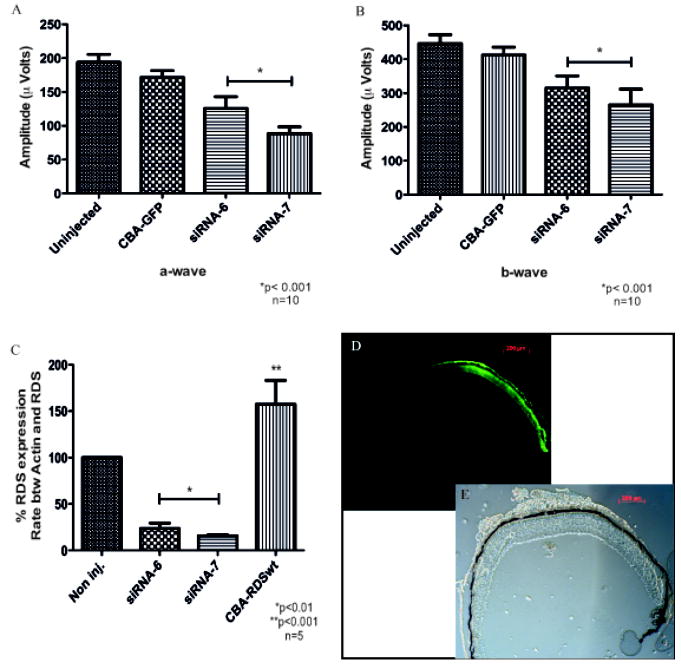
Fig. 29.2.
Reduction of rds in mice by siRNA delivered by rAAV to photoreceptors. Functional analysis by ERG: graphics show maximal amplitude of a-wave (A) and b-wave (B). Analysis of rds expression in the presence of siRNA 6 or 7 (C): total RNA was extracted and analyzed using real-time PCR. Transverse section of the retina showing its intact structure thought bright field (D) and a fluorescent picture of the same field (E) showing GFP expression in approximately ½ of the retina, corresponding to rAAV transduced area.

