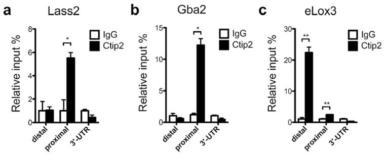
Figure 4. ChIP analyses on murine keratinocytes for lipid metabolizing genes.

Chromatin immunoprecipitation (ChIP) assay was performed on freshly isolated neonatal mouse epidermal keratinocytes using anti-Ctip2 antibody and results were analyzed by qPCR using primers indicated in table S2. Rat IgG was used as the control. Ctip2 was recruited to the promoter (relative to transcription start site) regions of (a) Lass2, (b) Gba2 and (c) eLox3. Statistical analyses were performed by student’s unpaired t-test using Graphpad Prism software. *p<0.05; **p<0.005. The results represent three separate experiments performed in triplicate.
