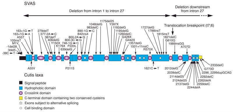
Figure 2. Schematic representation of the elastin domain organization.
The polypeptide consists of modules as defined on the lower left. The C-terminal region has also been described as a cell binding domain (74). Positions of mutations in the autosomal dominant cutis laxa (below) and in supravalvular aortic stenosis (SVAS) (above the molecule) are indicated by arrows.

