
Figure 1. Clinical and microscopic features of nevus sebaceus.
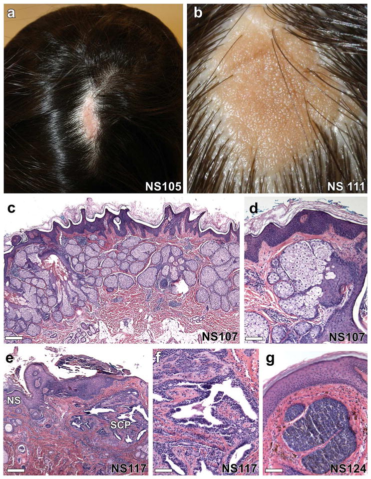
(a, b) Solitary, well-demarcated lesions on the scalp of two individuals show alopecia and a yellow-orange waxy appearance. (c) On histological examination, there is epidermal acanthosis, papillomatosis, and hyperkeratosis with dramatic increase in the number of sebaceous lobules and abortive hair follicles, scale = 288 μm, which is more evident at higher magnification (d), scale = 85 μm. (e–g) Up to 20% of nevus sebaceus lesions develop tumors including syringocystadenomas, trichoblastomas, trichilemmomas and tubular apocrine adenomas. (e) Nevus sebaceus (NS) with syringocystadenoma papilliferum (SCP) composed of villous structures lined by a columnar epithelium with stromal plasma cells, scale = 570 μm, most evident at higher magnification, (f), scale = 92 μm. (g) A trichoblastoma arising within a nevus sebaceus shows a well-circumscribed nodule of basaloid cells with a dense fibrocytic stroma, scale = 92 μm.