
Figure 2.
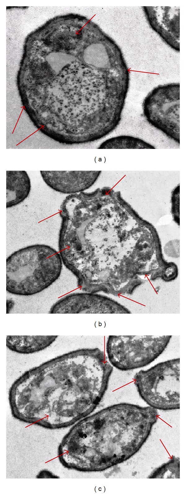
Transmission electron micrographs of untreated and treated S. Enteritidis cells. (a) Untreated cells having a regular outlined cell wall, plasma lemma lying closely to the cell wall, and regularly distributed cytoplasm (shown by arrows). (b) Methanolic P. arboris-vitae extract treated cells having extensive internal damage, unsymmetrical distributed cytoplasm, and larger and irregular periplasmic space (shown by arrows). (c) Ethanolic P. arboris-vitae extract treated cells having variable cell wall thickness appeared disrupted and variable periplasmic space (shown by arrows).