
Figure 2.
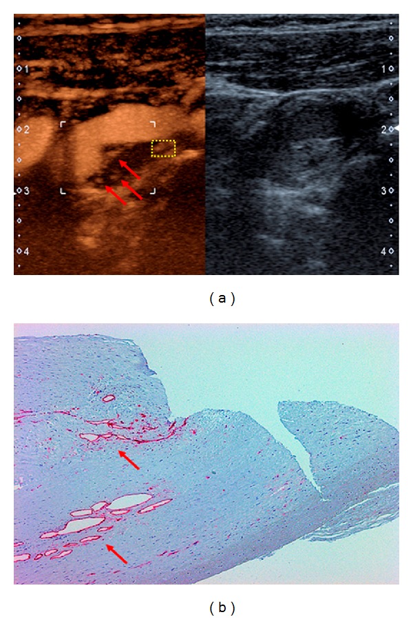
(a) Contrast-enhanced ultrasound (CEUS) of the carotid segment shown in Figure 1(a) (left). Intraplaque neovascularization is marked by red arrows. B-mode ultrasound of the same region (right). (b) Immunohistochemistry of CEA specimen corresponding to yellow-boxed area in (a). CD3 staining (red arrows) indicates plaque neovascularization.