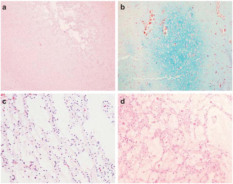
Figure 3.
Microcystic nodules (a) are characteristic of dysembryoplastic neuroepithelial tumors and demonstrate myxoid degeneration (b). The specific glioneuronal element is characterized by columns of OLCs between microcysts (c) and neurons that ‘float’ within the microcysts (d). (a, c, d: hematoxylin & eosin staining; b: alcian blue and periodic acid Schiff staining)

