
Figure 2.
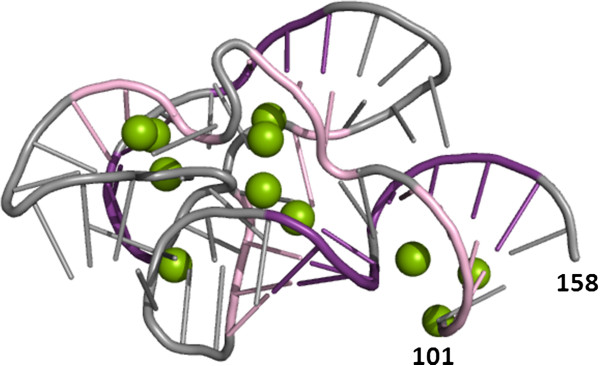
A traditional 2D rendering of the 3D structure for the 23S rRNA fragment (PDB ID: 1HC8, chain C). 5’ and 3’-terminal residues are numbered. Nucleotide residues involved in Watson-Crick base-pairing are shown in pink and violet. Mg2+ ions are shown as green balls. The limitation of this representation is that a single atom can have only one color and the use of multiple colors to illustrate different types of interactions can become overwhelming.