
Figure 7.
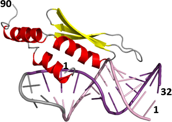
Protein-nucleic acid complex. A double-stranded RNA binding domain of S. cerevisiae RNAse III in complex with an AAGU tetraloop hairpin (PDB code: 2LBS, only the first model of the NMR ensemble is shown). The protein chain is represented as symbolic secondary structure cartoons: helices in red, strands in yellow. RNA molecule is represented as bonding sticks and colored according to secondary structure (base-paired residues are shown in violet and pink). Terminal residues are labeled.