
Figure 9.
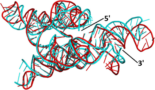
Comparison of a group I intron crystal structure and a comparative 3D model built with the ModeRNA program, in 3D. The homology model is compared to the crystal structure (1U6B_B), which was transformed by the deletion of 14 nt fragment to match the target sequence without major gaps. The picture shows both 3D structures aligned: crystal structure (cyan) and the model (red).