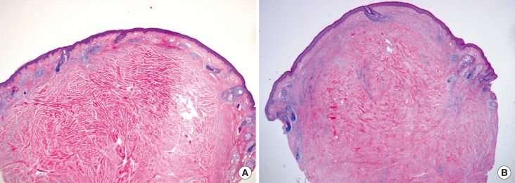
Fig. 3.

(A) First pregnancy. Histopathologic photograph showing thepresence of keloidal collagen and a prominent fascia-like fibrous band, which confirms the diagnosis of keloid (H&E, ×100). (B) Second pregnancy. Histopathologic photograph showing nearly the same appearanceof keloidal collagen and a prominent fascia-like fibrous band,as in the first pregnancy (H&E, ×100).
