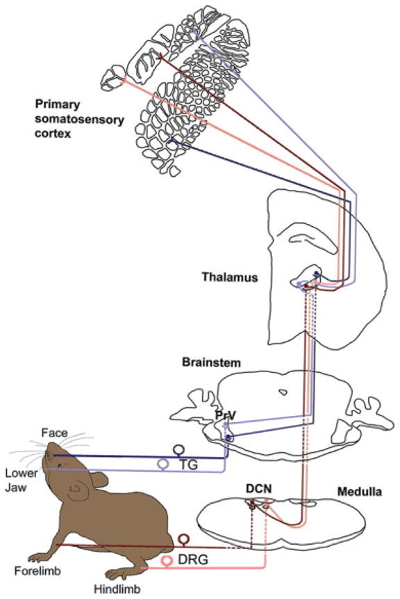
Fig. 1.

Mouse somatosensory pathways. Trigeminal ganglion–trigeminal lemniscal pathway (marked with blue) carries the orofacial inputs to the VPM and subsequently to the facial areas of the primary somatosensory (SI) cortex. The pathway schematized by dark blue color carries sensory information from the face; the pathway marked with light blue carries information from the lower jaw. Dorsal root ganglion– dorsal column lemniscal pathway is marked in red. Sensory information from hindlimb (light red) and forelimb (dark red) are first transmitted to the dorsal column nuclei (gracile nucleus and cuneate nucleus, respectively), then to the contralateral VPL, and then to the paw representation areas of the SI cortex.
