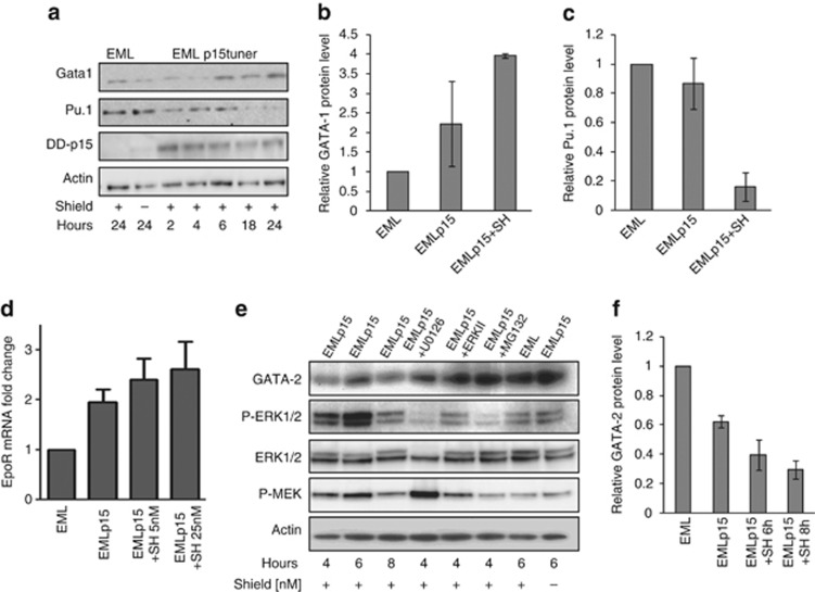
Figure 7.
Molecular changes following the expression of p15Ink4b in EML cells. (a) Time-dependent changes in GATA-1, Pu.1 and p15Ink4b protein expression following the addition of SH (100 nℳ) to induce accumulation of p15Ink4b protein in EMLp15Tuner cells (EMLp15). EML cells treated with SH or untreated EMLp15Tuner cells, harvested at 24 h, were used as controls (n=3). (b) Increase in GATA-1 protein levels at 24 h following the addition of SH. (c) Decrease in Pu.1 protein levels at 24 h following the addition of SH. Blots were quantitated using ImageJ and normalized to actin (n=2). (d) EpoR mRNA expression in EMLp15Tuner cells following 24 h exposure to SH (5 and 25 nℳ). Expression was normalized to 18SrRNA and depicted as fold change of the mRNA levels in EML cells (n=3). (e) Time-dependent changes in GATA-2 expression and phosphorylation of MEK and ERK1/2 proteins following the addition of SH (100 nℳ).The observed decrease in GATA-2 is abolished by the addition of MEK1/2 inhibitor (U0126), ERK inhibitor (ERKII) or 26S proteasome inhibitor (MG132). EML cells treated with SH or untreated EMLp15Tuner cells, harvested at 6 h, were used as controls. (f) Relative decrease in GATA-2 protein levels at 6 and 8 h following the addition of SH. Blots were quantitated using ImageJ and normalized to actin (n=2).

