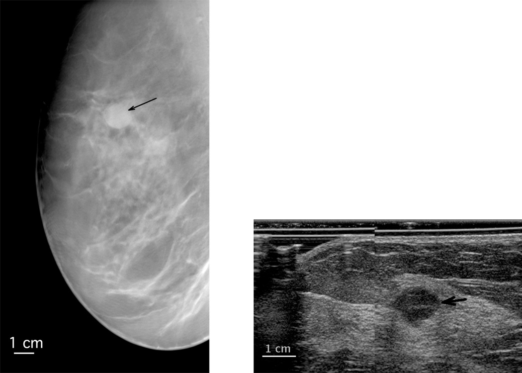
Figure 2.
Sections extracted from the LMO views of a metaplastic carcinoma in a right breast (left DBT, right 3D-AUS, the black arrow points toward the mass), diagnosed as benign on DBT by three readers. The AUS image is from a plane normal to the image plane of the DBT at the level of the white arrows on the DBT. The ultrasound is magnified relative to the DBT.

