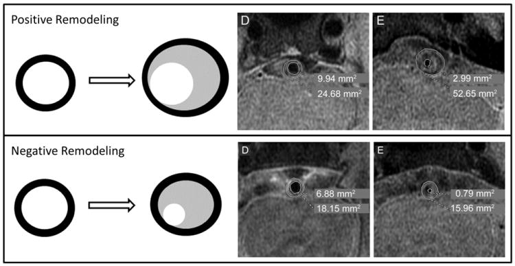
Figure 1. Illustration of positive (outward) and negative (inward) remodeling in ICAD.

Positive Remodeling: Top panel (left) shows diagram of positive remodeling: the vessel wall (black) expands outward with accumulation of lipid (gray) and may or may not decrease the lumen (white). Top panel (right) shows an example of positive remodeling in a case of severe basilar stenosis (reproduced from Ma et al. Neurology, 201020). At the level of maximal stenosis (E), the wall area is increased and the lumen area is decreased relative to a distal basilar segment (D). Negative remodeling: Bottom panel (left) shows diagram of negative remodeling: the vessel wall (black) does not expand outward with accumulation of lipid (gray) and results in decrease in the lumen (white). Bottom right panel shows an example of negative remodeling in a case of severe basilar stenosis (reproduced from Ma et al. Neurology, 201020). At the level of maximal stenosis (E), the wall area is slightly decreased and the lumen is decreased relative to a distal basilar segment (D).
