
Fig. 10.
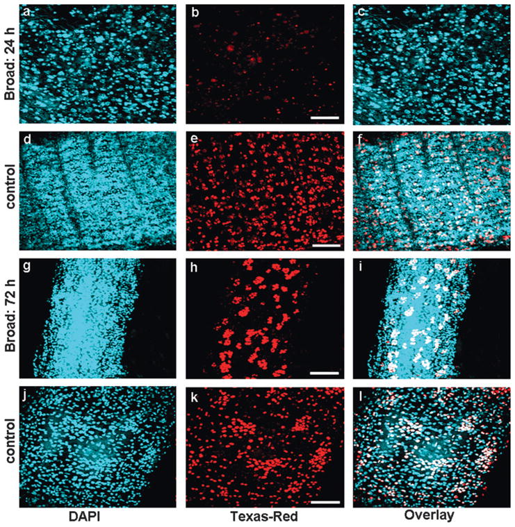
Effect of Tcbr RNAi on the cell proliferation of midgut. Cell proliferation assay was performed using BrdU. BrdU was administered in insects injected with Tcbr or malE (control) dsRNA after four days of injection. The detection of proliferating cells was performed using anti-BrdU primary antibody and Texas-Red conjugated secondary antibody. Panels show nuclear staining by DAPI (a, d, g and j), proliferating cells staining by BrdU (b, e, h and k) and overlay of both (c, f, i and l). Injection of Tcbr dsRNA at 24 AEFL blocked cell proliferation in midgut and only a few BrdU positive cells were observed (b) when compared to its control (e). Injection of Tcbr dsRNA at 72 h AEFL showed midgut in advanced state of proliferation with evagination of crypts (h) when compared to its control (k). Only proliferating imaginal cells were detected by BrdU (white in overlay images); larval or differentiated pupal cells remain unstained (blue in overlay images). Controls include insects not injected with BrdU (not shown). Scale bar: 20 μm.