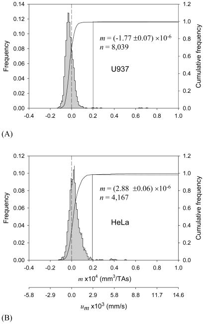
Figure 1.
Magnetophoretic mobility (MM, m) histograms of (A) U-937 cells (24-hour incubation in complete media, viability 90.9%), and (B) HeLa cells (19-hour incubation, viability 96.1%). The second axis at the bottom shows the corresponding magnetic field-induced velocity (um) values. Drop lines indicate cut-off mobility used to discriminate between “high” and “low” MM fractions (see text).

