
Figure 2.
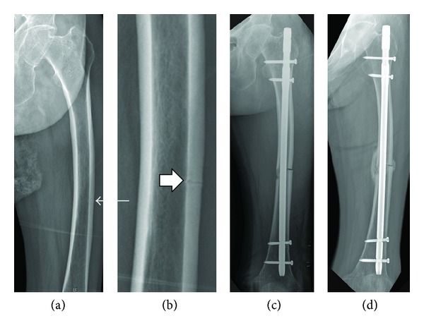
(a) A 74-year-old woman suffered thigh pain and her AP radiograph showed exaggerated femoral bowing with a transverse radiolucent line (arrow) in the lateral cortex of the distal 1/3 of the left femur. (b) A magnified view (box arrow) of the lesion revealed an incomplete, prefracture lesion of an atypical femoral fracture. (c) After preventive IM nailing, a complete fracture occurred. (d) The union was achieved with callus bridging at 18 weeks postoperatively.