
Figure 3.
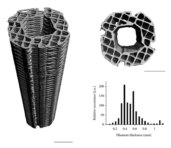
Side and top view of a PCL scaffold for tibia segmental defect regeneration, visualised by microcomputed tomography. The fabrication technique results in scaffolds with well-controlled architecture as evidenced by the narrow filament thickness distribution, leading to a porosity (volume fraction available for tissue ingrowth) of 60%, with interconnected pores. Scale bars are 5 mm.