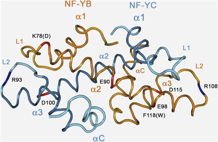
Figure 6.
Structure of the Human NF-Y HFD Subunit Dimer.
Ribbon representation of human NF-YB/NF-YC dimer structure (Protein Data Bank code: 1N1J) and location of selected residues, determined by Romier et al. (2003). NF-YB and NF-YC main chains traces are represented in orange and cyan color, respectively. Secondary structure elements are labeled, and main chain location of selected relevant residues described in the text (human numbering) is highlighted in blue (positive) or red (negative) for side-chain charge. NF-YB Lys-78 (Asp in LEC1/L1L proteins) is colored in red. Human NF-YB Phe-118 (Trp in plants) is highlighted in green.

