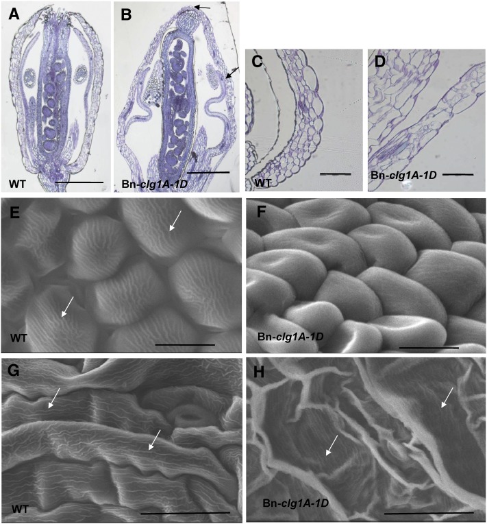
Figure 8.
Changes in Flower Morphology and Surface Characteristics of Arabidopsis Plants Transformed with Bn-clg1A-1D as Observed by Light and Electron Microscopy.
(A) to (D) Light microscopy images of Toluidine Blue–stained longitudinal sections of flowers at developmental stage 12 ([A] and [B]) and sepals at developmental stage 13 ([C] and [D]). Col-0 wild-type (WT) ([A] and [C]) and transgenic Bn-clg1A-1D plants ([B] and [D]) are represented. Arrows indicate sepal-to-sepal and sepal-to-petal fusions.
(E) to (H) Scanning electron microscopy images of abaxial surfaces of petal ([E] and [F]) and sepal ([G] and [H]) epidermis in Col-0 wild-type ([E] and [G]) and transgenic Bn-clg1A-1D plants ([F] and [H]). Arrows indicate nanoridges.
Bars = 500 µm in (A) and (B), 100 µm in (C) and (D), 10 µm in (E) and (F), and 30 µm in (G) and (H).
[See online article for color version of this figure.]

