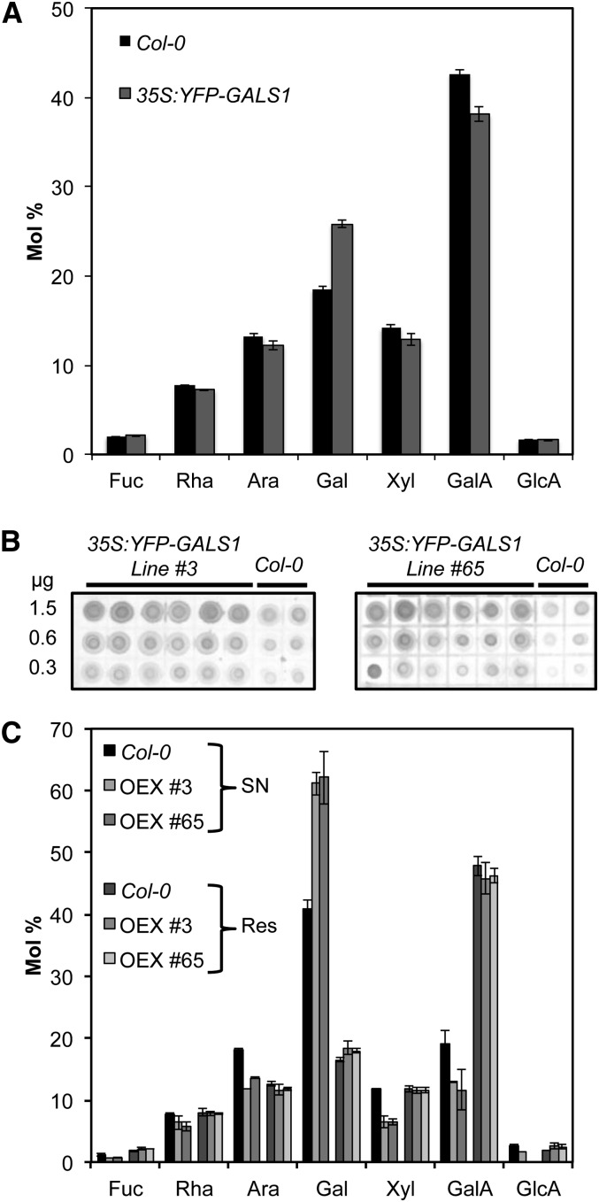
Figure 7.
Characterization of Cell Walls in Plants Overexpressing GALS1.
(A) Cell wall monosaccharide composition of leaves from 4-week-old Arabidopsis Col-0 wild-type plants and overexpressing plants transformed with 35Spro:YFP-GALS1. Values are means ± sd obtained with six wild-type plants and seven T2 plants from lines #3 and #65. The cell wall Gal content in the overexpressors was significantly increased by ∼50% (ANOVA, P < 0.0001), whereas the ratio between other monosaccharides was not significantly changed.
(B) Immunodot blot analysis of leaf cell walls from 4-week-old T2 plants from two different T1 lines transformed with 35Spro:YFP-GALS1. The blots were developed with the LM5 monoclonal antibody and show increased content of β-1,4-galactan in the overexpressors. Values on the left indicate the amount of cell wall material spotted on each line of dots.
(C) Leaf cell walls from 4-week-old wild-type and YFP-GALS1 overexpressing plants were digested with endo-β-1,4-galactanase, separated into supernatant (SN) and residue (Res), and analyzed by HPAEC. The digestion released 21.2% ± 1.8% of total Gal in the wild type and 36.8% ± 0.3% in the overexpressors (OEX). Data show means ± sd (n = 4). Gal content was significantly different between overexpressors and the wild type in the supernatant (t test, P < 0.003) but not in the residue.

