Fig. 2.
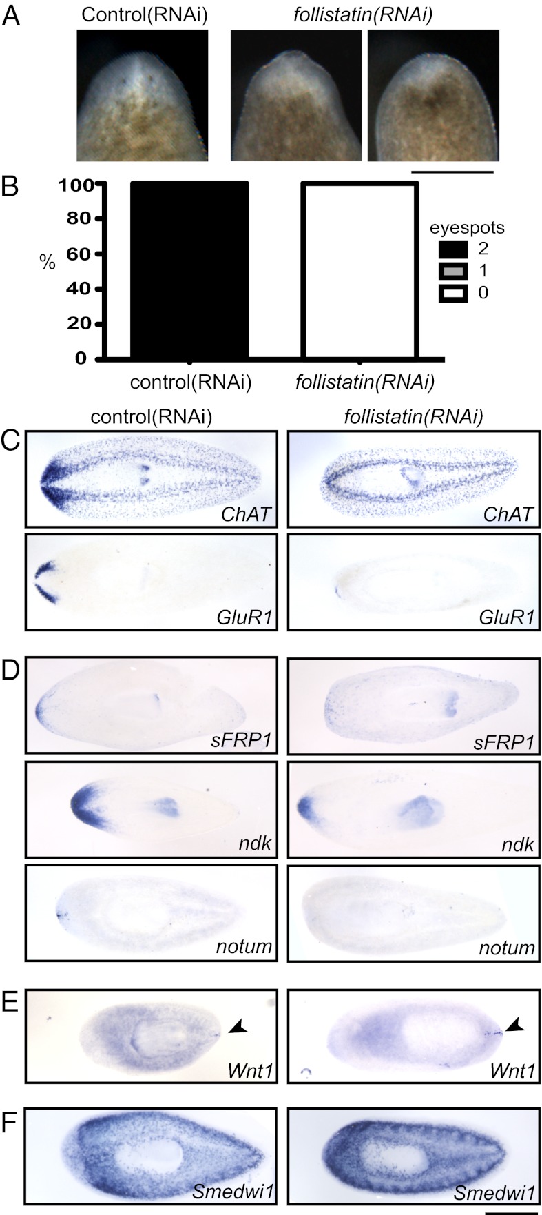
follistatin plays a critical role in anterior regeneration. (A) After 5 d of head regeneration, follistatin(RNAi) animals display small blastemas compared with control animals and are missing eyespots. (B) Nearly all control animals regenerated eyespots within 5 d of amputation, but eyespots were missing from all follistatin(RNAi) animals (n ≥ 50 each). (C) Cephalic ganglia are absent or dramatically reduced in size in follistatin(RNAi) animals 5 d after amputation of the head. Both control(RNAi) and follistatin(RNAi) animals were subjected to in situ hybridization with ChAT and GluR1 probes to mark the entire central nervous system and the brain branches, respectively. (D) Anterior marker expression was reduced after 5 d of head regeneration in follistatin(RNAi) animals. sFRP-1, ndk, and notum probes each mark different anterior cell populations. (E) A posterior marker, wnt1, was expressed in an expanded posterior region (arrowheads) but was not expressed inappropriately in the anterior of follistatin(RNAi) animals. (F) In situ hybridization with a probe for a neoblast marker, Smedwi-1, indicates that neoblasts were present after follistatin(RNAi). (Scale bars, 500 µm.) Anterior is up (A) or to the left (C–F).

