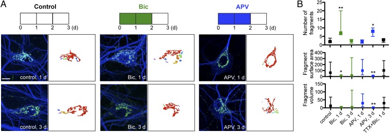
Fig. 2.
Prolonged treatment with bicuculline (20 μM) or removal of APV (200 μM DL-APV) fragments the Golgi complex. (A) Immunostaining of cultured hippocampal neurons (≥21 DIV) with cis-Golgi marker anti-GM130 (green) and anti-MAP2 (blue) with 3D reconstruction of Golgi staining. (Scale bar: 10 μm.) (B) Quantification of number, surface area (μm2), and volume (μm3) of distinct Golgi fragments from reconstructed anti-GM130 fluorescent signal. Application of TTX (1 μM) before bicuculline to block synaptic transmission. Data shown are median and IR (control, n = 10; Bic, 1 d, n = 7; Bic, 3 d, n = 6; APV, 1 d, n = 9; APV, 3d, n = 7; TTX+Bic, 1 d, n = 5). For Bic, 1 d, P < 0.1.

