Fig. 4.
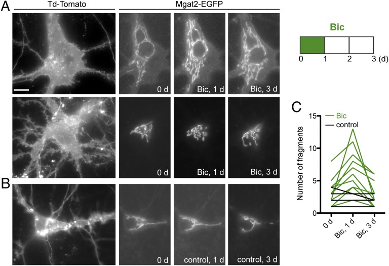
Golgi fragmentation occurs during bicuculline treatment and reverses after return to normal medium. Cultured hippocampal neurons were transfected with Mgat2–EGFP and myristoylated Td-Tomato. Individual neurons were imaged, then treated with bicuculline for 1 d. Bicuculline was removed and neurons were imaged again after 2 d in normal medium. (A) Examples of two neurons with fragmentation of the Golgi complex after 1 d with bicuculline, then reversal of fragmentation 2 d after bicuculline removal. (Scale bar: 15 μm.) (B) Example control neuron showing change in Mgat2–EGFP signal but lack of fragmentation. (C) Summary of data from bicuculline-treated (green, n = 12) and control (black, n = 4) neurons.

